|
|
楼主 |
发表于 2008-8-22 01:32:14
|
显示全部楼层
英格力士版(部分翻译在5楼):
Heterochromia
In anatomy, heterochromia refers to a difference in coloration, usually of the iris but also of hair or skin. Heterochromia is a result of the relative excess or lack of melanin (a pigment). It may be inherited, or due to genetic mosaicism, or due to disease or injury.
Eye color, specifically the color of the irides, is determined primarily by the concentration and distribution of melanin. The affected eye may be hyperpigmented (hyperchromic) or hypopigmented (hypochromic).In humans, usually, an excess of melanin indicates hyperplasia of the iris tissues, whereas a lack of melanin indicates hypoplasia.
Heterochromia of the eye (heterochromia iridis or heterochromia iridum) is of two kinds. In complete heterochromia, one iris is a different color from the other. In partial heterochromia or sectoral heterochromia, part of one iris is a different color from its remainder.
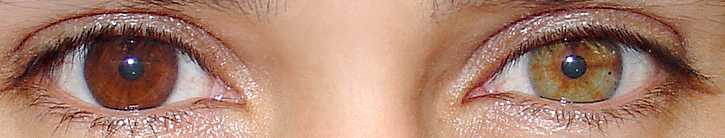
Complete heterochromia in human eyes: one brown and one hazel
Partial or sectoral heterochromia is much less common than complete heterochromia and is typically found in autosomally inherited disorders such as Hirschsprung's disease and Waardenburg syndrome. Famous comedian Dan Aykroyd has heterochromia, as do singer/songwriter Carly Simon, actresses Kate Bosworth, Elizabeth Berkley, Mila Kunis, Jane Seymour, Eleni Willmott, actor Christopher Walken, American mixed martial artist Jens Pulver, Rock singer Tim McIlrath, and Major League Baseball pitcher Max Scherzer. Musician David Bowie is often thought to have heterochromia, but this is not the case as Bowie's eyes are both blue (his left pupil is permanently dilated due to a childhood injury).

Sectoral heterochromia: a blue iris with a brown section.
Classification based on etiology
Heterochromia is classified primarily by onset: as either genetic or acquired. Although a distinction is frequently made between heterochromia that affects an eye completely or only partially (sectoral heterochromia), it is often classified as either genetic (due to mosaicism or congenital) or acquired, with mention as to whether the affected iris or portion of the iris is darker or lighter.
Congenital heterochromia
Heterochromia that is congenital is usually inherited as an autosomal dominant trait.
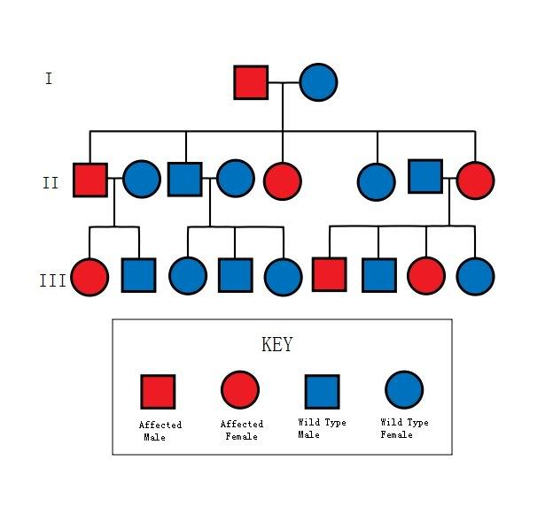
Congenital heterochromia: inherited in autosomal dominant fashion (from men or women).
Abnormal iris darker
* Lisch nodules — iris hamartomas seen in neurofibromatosis.
* Ocular melanosis — a condition characterized by increased pigmentation of the uveal tract, episclera, and anterior chamber angle.
* Oculodermal melanocytosis (nevus of Ota)
* Pigment dispersion syndrome — a condition characterized by loss of pigmentation from the posterior iris surface which is disseminated intraocularly and deposited on various intraocular structures, including the anterior surface of the iris.
* Sturge-Weber syndrome — a syndrome characterized by a port-wine stain nevus in the distribution of the trigeminal nerve, homolateral meningeal angioma with intracranial calcification and neurologic signs, and angioma of the choroid, often with secondary glaucoma.
Abnormal iris lighter
* Simple heterochromia — a rare condition characterized by the absence of other ocular or systemic problems. The lighter eye is typically regarded as the affected eye as it usually shows iris hypoplasia. It may affect an iris completely or only partially.
* Congenital Horner's syndrome — sometimes inherited, although usually acquired
* Waardenburg's syndrome — a syndrome in which heterochromia presents as a bilateral iris hypochromia in some cases. A Japanese review of 11 albino children with the disorder found that all had sectoral/partial heterochromia.
* Piebaldism — similar to Waardenburg's syndrome, a rare disorder of melanocyte development characterized by a white forelock and multiple symmetrical hypopigmented or depigmented macules.
* Hirschsprung's disease — a bowel disorder associated with heterochromia in the form of a sector hypochromia. The affected sectors have been shown to have reduced numbers of melanocytes and decreased stromal pigmentation.
* Incontinentia pigmenti
* Parry-Romberg syndrome
Acquired heterochromia
Heterochromia that is acquired is usually due to injury, inflammation, the use of certain eyedrops, or tumors.
Abnormal iris darker
* Deposition of material
a) Siderosis — iron deposition within ocular tissues due to a penetrating injury and a retained iron-containing, intraocular foreign body.
b) Hemosiderosis — long standing hyphema (blood in the anterior chamber) following blunt trauma to the eye may lead to iron deposition from blood products
* Use of certain eyedrops — prostaglandin analogues (latanoprost, isopropyl unoprostone, travoprost, and bimatoprost) are used topically to lower intraocular pressure in glaucoma patients. A concentric heterochromia has developed in some patients applying these drugs. The stroma around the iris sphincter muscle becomes darker than the peripheral stroma. A stimulation of melanin synthesis within iris melanocytes has been postulated.
* Neoplasm — Nevi and melanomatous tumors.
* Iridocorneal endothelium syndrome
* Iris ectropion syndrome
Abnormal iris lighter
* Fuchs' heterochromic iridocyclitis — a condition characterized by a low grade, asymptomatic uveitis in which the iris in the affected eye becomes hypochromic and has a washed-out, somewhat moth eaten appearance. The heterochromia can be very subtle, especially in patients with lighter colored irides. It is often most easily seen in daylight. The prevalence of heterochromia associated with Fuch's has been estimated in various studies with results suggesting that there is more difficulty recognizing iris color changes in dark-eyed individuals.
* Acquired Horner's syndrome — usually acquired, as in neuroblastoma, although sometimes inherited.
* Neoplasm — Melanomas can also be very lightly pigmented, and a lighter colored iris may be a rare manifestation of metastatic disease to the eye.
Heterochromia has also been observed in those with Duane syndrome.
* Chronic iritis
* Juvenile xanthogranuloma
* Leukemia and lymphoma
Central heterochromia

A grey-blue iris with a greenish-yellow ring showing Central Heterochromia

A grey iris featuring Central Heterochromia
Whereas Heterochromia (also known as a heterochromia iridis or heterochromia iridum) is an eye condition in which one iris is a different colour from the other (complete heterochromia), Central Heterochromia is an eye condition in which there are two different colours in the same iris. Central Heterochromia is where the central (pupillary) zone of the iris is a different colour than the mid-peripheral (ciliary) zone.
Eye colour is determined primarily by the concentration and distribution of melanin pigment within the iris tissues, Anything affecting those factors may result in a difference of colour being observed.
The human iris can be seen in a number of various colours. There are three true colors in the eyes that determine the outward appearance; brown, yellow, and grey. How much of each colour an individual has determines the appearance of his or her eye colour.
Eyes displaying Central Heterochromia are often referred to as "cat eyes" because of the appearance of a multi-coloured iris. Central Heterochromia appears to be prevalent in irides containing low amounts of melanin.Central Heterochromia does not label an eye as hazel. This is because the outer ring of an eye affected by Central Heterochromia is that iris' true colour.
[ 本帖最后由 踏歌行 于 2008-8-28 21:11 编辑 ]
SO现金网╬足球.时时彩.快乐十分SO.CC
(六_合 )投注网╰倍率48.5倍1111.cc
性感╯美女bbs.334.cc |
|


 发表于 2008-8-22 01:31:34
 发表于 2008-8-22 01:33:10


